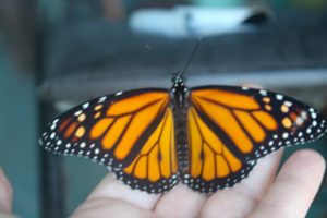
Many of us are distributing Monarch butterflies as a means to excite the public, especially schoolchildren, about the wonders of butterflies, metamorphosis, and migration. Unfortunately, there is a good chance that our efforts may be actually harming Monarchs by increasing the frequency of an insidious parasite. We have experienced this parasite in our rearing operations at the Universities of Minnesota and Kansas, and know that we have inadvertently distributed larvae that may have been infected. Since most people that receive Monarchs release them, this is likely to result in a higher frequency of infected butterflies. We have been working for the past year on controlling this parasite and would like to share our results with you in the hope of preventing further negative effects on an organism that we all view with a sense of awe and wonder.
The parasite is a protozoan, with the Latin name of Ophryocystis elektroscirrha. It was first described over 20 years ago but only began receiving a great deal of attention in the last several years. The parasite begins its life cycle as an inactive spore that needs to be eaten by a larva. It multiplies within the larva, and during the last few days of the pupal stage produces new spores that are on the butterfly’s scales when it emerges. It is then transferred to the surface of the egg or milkweed during oviposition and begins a new cycle when it is eaten by the emerging larva. We have shown that the spores can be transferred from an infected male to a healthy female during mating and that this female can transfer them to her offspring. Spores can also be transferred passively when an infected butterfly is held in a cage with healthy butterflies (and probably also in the dense overwintering clusters in California and Mexico), although we have not yet learned whether this means of spore transfer results in infected offspring from an otherwise healthy female.
You may have experienced this parasite. If you have noticed pupae with dark gray blotches on them just before you see the butterfly through the pupa covering, or if some of your butterflies fail to emerge completely and seem stuck within the pupa, it is probably because they are infected. This is more likely to happen if you rear more than one generation using the offspring from one generation as parents of the next, or if you use the same cages for several generations. However, these symptoms only appear with very high levels of infection, and your pupae and adults can appear perfectly normal and still be heavily infected.
What should be done? We have worked out a way to control this parasite that we hope will not be too difficult. Our method requires cleaning up your rearing operation; we have not yet found a way to “cure” a larva once it has eaten the spores, although at the University of Kansas we are continuing to look for such a solution using drugs that have been shown to work on related organisms. We have had limited success with attempts to surface-decontaminate eggs once they have been laid, although this does lower the incidence of the disease. Thus, the only way to solve the problem, and to prevent more releases of contaminated butterflies, is to make sure that the larvae you rear are never exposed to the parasite. There are four steps you will need to take:
1. Clean all of your rearing cages and other equipment. It is difficult to clean wooden cages unless you have access to an autoclave. At Minnesota, we use wood and screen cages to rear larvae and have successfully decontaminated them in an autoclave. If you use plastic cages, they can be decontaminated by soaking them in a 10% bleach solution (approximately 10 ml Chlorox bleach to 100 ml water) or 100% ethanol for at least 15 minutes, then rinsed well. Use the bleach solution to soak any tools that you use to transfer larvae, rinsing them after they are soaked. Wipe down countertops and other surfaces with the bleach solution in areas in which you have reared larvae or kept butterflies. The spores survive long periods of time (over a year), and can also survive freezing temperatures, so equipment that you used last year or left outside over the winter will still be able to infect larvae.
2. Check all females you use to produce eggs to make sure that they are not contaminated with spores. This is especially important if they have been reared in captivity, but wild females should also be checked. To look for spores, you will need tweezers or forceps, clear scotch tape (not the “magic”, slightly foggy kind), microscope slides, and access to a microscope. Your local high school, veterinary clinic, or the biology department of a university or college might have microscopes that you could use.
a. Cut a small piece of tape, handling it with the tweezers so your fingerprints aren’t on it.
b. Hold the butterfly’s wings in your left hand (if you’re right-handed) so that the abdomen is exposed. Holding the tape with tweezers in your right hand, carefully use it to rub off a patch of scales from the end of the butterfly’s abdomen. If you are doing several butterflies, wash your hands and sterilize the tweezers often. Put the tape, sticky side down, on the microscope slide. You can put 3 or 4 pieces of tape on the same slide. Write identifying numbers next to the tape pieces so that you’ll know which results go with which butterfly.
c. Scan the entire piece of tape for spores under 100-200x magnification. These are amber colored, and are much smaller than scales (see attached photograph). They are football-shaped, and have a very regular, or constant, appearance. With practice you can detect the spores under a dissecting scope, but we recommend using a compound microscope when you are first learning to recognize the spores. A badly infected individual will have thousands of spores, but often there will be only a few. They often occur in clumps of several spores between the scales, and sometimes they are on the surface of the scales.
d. If you see any spores, kill the infected butterfly and dispose of it. This can be done by quickly cutting off its head, or putting it in an envelope and freezing it.
e. If you do not have access to a microscope or microscope slides, we will provide slides and check them for spores. Please contact Karen Oberhauser (address below) if you would like this help.
3. If your females have no spores, they can be used to obtain eggs. If you mate them, check the males too.
4. You should go through an entire generation before disseminating any larvae or pupae to people that might release them. This step is important if you have used your present equipment, or even rearing space, before, especially if you have experienced the symptoms described above. Since it is impossible to tell if a larva or pupa is infected without dissecting it, you should rear all of this generation to adulthood, and check the butterflies as soon as they can be handled. If fewer than 5 out of every 100 adults are infected, it should be safe to distribute larvae and pupae from the next generation. Be sure, however, to continue to follow steps 1 through 3. Infected butterflies should all be killed.
It took us two generations to completely get rid of the disease. In Minnesota, we are continuing to study the disease and purposely inoculate some eggs with spores so that we can learn more about how it is transmitted. Even though this requires that there are some spores around, by being very careful to keep infected stock away (this means in a different room) from healthy stock and sterilizing all equipment, we have experienced no infected individuals in uninoculated stock for two generations. We continue to check every butterfly that emerges for spores, however.
We realize that this might seem burdensome, especially if you sell Monarchs. It is also hard and seems cruel, to kill butterflies that look perfectly healthy. We do not see an alternative, however, and believe that all of us share a common interest and responsibility to preserve the health of this amazing organism.


